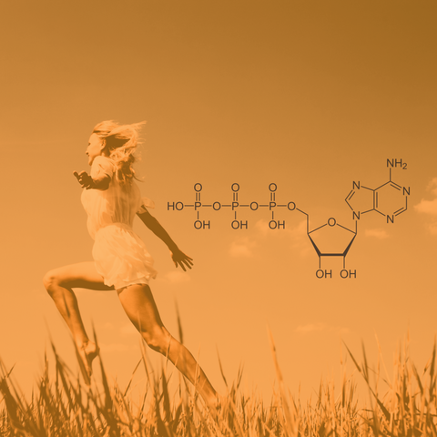
The mitochondria are crucial and ubiquitous organelles in eukaryotic cells whose main function is to generate energy in the form of adenosine triphosphate (ATP) through the oxidative phosphorylation system (OXPHOS). OXPHOS is composed of the electron transport chain, formed by four multimeric enzymes and two mobile electron carriers, that performs the redox oxidations involved in cellular respiration and generates the proton motive force required to create ATP [1].
Most electrons enter into the respiratory chain with NADH-ubiquinone oxidoreductase (or complex I) before reaching succinate dehydrogenase (complex II), ubiquinol–cytochrome c oxidoreductase (complex III), and cytochrome c oxidase (complex IV). The last respiratory chain component, called ATP synthase (complex V) [2], is responsible for the conversion of adenosine diphosphate (ADP) and inorganic phosphate (pi) into ATP [3,4].
ATP synthase (also referred to as F1 Fo -ATPase or H+ -ATPase) is an extraordinarily prevalent complex of proteins found in all living organisms. Despite variations, ATP synthases are highly similar across species both structurally and functionally. In bacteria, the enzyme is found in the cytoplasmic membrane and provides ATP necessary for its biosynthesis, transport, or motility. In plants, ATP synthase, found in the thylakoid membranes of chloroplasts, is involved in photosynthesis. In animals, ATP synthase is located in the inner membrane of mitochondria and the cytoplasmic membranes of various cells, such as endothelial cells, keratinocytes, and adipocytes [4].
ATP synthase, known as one of the smallest biological nanomotors, produces an estimated total of about 2.0 million kg of ATP during the 75-year lifespan of a human being [4]. The number of protons required to synthesize one molecule of ATP varies from three to four among different organisms, with the possibility for cells to change this ratio while adapting to their environmental conditions [5,6]. Finally, recent studies support previously published estimates that ATP synthase operates with an exceptional efficiency rate of approximately 90% [7].
The structure of ATP synthase in humans
The human mitochondrial ATP synthase is a protein complex composed of multiple subunits, collectively estimated to have a molecular weight of 550 kDa. Some of these subunits are associated with the inner mitochondrial membrane [8]. In total, this complex comprises 29 polypeptide chains of 18 subunits and is composed of two main domains, the Fo and F1 domains. The Fo domain (consisting of c8-ring, ATP6, or a, ATP8 or A6L, e, f, g, DAPIT, and 6.8 PL in the inner mitochondrial membrane) is responsible for proton translocation. The F1 domain (the soluble head consisting of the α3β3 subcomplex) is responsible for the synthesis of ATP. Both domains are linked by a central stalk (γ, δ, and ε subunits) that can rotate inside the F1 domain and a peripheral stalk (F6, b, d, and OSCP subunits) that remains stationary [8-10].
Theligomerization of ATP synthase in dimers is essential for enhancing its activity and producing energy by establishing and preserving local proton charge and mitochondrial membrane potential [11]. Its organization into dimers is also crucial for the formation of the invaginations of the inner mitochondrial membrane. ATP synthases shape the cristae membrane and may play a beneficial physiological role [12]. In human mitochondria, ATP synthase is associated with the ATPase inhibitory factor 1 (IF1) protein. IF1 can inhibit the ATP hydrolytic activity of ATP synthase during its assembly or under certain conditions, such as a collapse of the proton motive force [13-15].
The role and function of ATP synthase in humans
ATP production
The mitochondrion has an outer membrane that encloses the organelle and an inner membrane with folds called cristae, creating compartments known as the lumen (internal space within the cristae of the inner mitochondrial membrane). The matrix is the central compartment enclosed by the inner mitochondrial membrane that contains essential enzymes (complexes I, II, III, and IV) for many biochemical reactions to generate energy in the form of ATP. Electron transfer along the respiratory chain is coupled to the movement of protons from the matrix to the cristae lumen, establishing an electrochemical proton gradient (or proton motive force) that drives ATP production by ATP synthase [16]. ATP synthase uses the energy derived from the electrochemical gradient to phosphorylate ATP from A [8].
The "binding change mechanism" is a fundamental component of the ATP synthase catalytic mechanism, involving rotational motion and steady-state catalysis [17]. The transport and catalytic activities of the ATP synthase complex rely on the functions of its subunits. A complete 360-degree turnover at each catalytic site involves achieving and transitioning through three conformations, and a cycle concludes with a 120-degree rotation at a separate catalytic site. The three catalytic sites (3α and 3β subunits forming the α3β3 subcomplex) of the enzyme bind ADP and inorganic phosphate in sequence before undergoing a conformational change to generate ATP. The sites then change conformation again allowing the release of ATP [18]. The enzyme's inner core undergoes rotation to facilitate these conformational changes, driven by the proton motive force generated by protons crossing the mitochondrial membrane. A circular pattern of alternating positions of 3α and 3β subunits, each exhibiting distinct conformations and functions, can be observed [19].
Cellular apoptosis induction
Apart from producing ATP, ATP synthase can also induce cellular apoptosis by creating a mitochondrial permeability transition pore that triggers programmed cell death upon opening. The resulting disruption of the electron transport chain causes depolarization, depletion of pyridine nucleotides, release of calcium, respiratory inhibition, as well as mitochondrial matrix swelling. This swelling can lead to the release of pro-apoptotic proteins like cytochrome c or endonuclease G, among others [8].
ATP synthase, a drug target?
ATP synthase is a vital enzyme in bacteria and therefore represents a potential target for antimicrobial agents. However, therapeutic applications in humans are constrained by the similarity of ATP synthases across species. Several antibiotics, such as efrapeptins, aurovertins, or oligomycin, inhibit its function [20]. For example, oligomycin binds to the Fo domain of ATP synthase and inhibits proton conduction in bacteria. However, its use in humans is impossible due to its toxic effects on mitochondrial ATP synthase.
Bedaquiline is the only ATP synthase inhibitor approved for the treatment of bacterial infections in humans, specifically for pulmonary multidrug-resistant tuberculosis [21-23]. As oligomycin, bedaquiline inhibits ATP synthases not only in bacteria but also in mitochondria, although it may not cause ATP synthesis-related toxicity in mammalian cells [24].
Conclusion
ATP synthase is a remarkable enzyme with profound implications for cellular energy production and various physiological processes. It is a universal energy converter that plays a critical role in cellular functions.
If you're looking to increase your ATP production and gain more energy, you can experience pure cellular revitalization with Just Blue, our pure 16 mg methylene blue buccal troche to brighten your focus, decrease inflammation, and boost your energy and endurance. Methylene blue enhances energy production by donating electrons to the electron transport chain, increasing ATP via the ATP synthase complex.
References
[1] Del Dotto, V., Musiani, F., Baracca, A. and Solaini, G. (2024) Variants in Human ATP Synthase Mitochondrial Genes: Biochemical Dysfunctions, Associated Diseases, and Therapies. International Journal of Molecular Sciences, 25, 2239. https://doi.org/10.3390/ijms25042239
[2] Sharma, L., Lu, J. and Bai, Y. (2009) Mitochondrial Respiratory Complex I: Structure, Function and Implication in Human Diseases. Current Medicinal Chemistry, 16, 1266–77. https://doi.org/10.2174/092986709787846578
[2] Sharma, L., Lu, J. and Bai, Y. (2009) Mitochondrial Respiratory Complex I: Structure, Function and Implication in Human Diseases. Current Medicinal Chemistry, 16, 1266–77. https://doi.org/10.2174/092986709787846578
[3] Zharova, T.V., Grivennikova, V.G. and Borisov, V.B. (2023) F1·Fo ATP Synthase/ATPase: Contemporary View on Unidirectional Catalysis. International Journal of Molecular Sciences, 24, 5417. https://doi.org/10.3390/ijms24065417
[4] Ahmad, Z., Okafor, F., Azim, S. and F. Laughlin, T. (2013) ATP Synthase: A Molecular Therapeutic Drug Target for Antimicrobial and Antitumor Peptides. Current Medicinal Chemistry, 20, 1956–73. https://doi.org/10.2174/0929867311320150003
[5] Van Walraven, H.S., Strotmann, H., Schwarz, O. and Rumberg, B. (1996) The H + /ATP coupling ratio of the ATP synthase from thiol‐modulated chloroplasts and two cyanobacterial strains is four. FEBS Letters, 379, 309–13. https://doi.org/10.1016/0014-5793(95)01536-1
[6] Yoshida, M., Muneyuki, E. and Hisabori, T. (2001) ATP synthase — a marvellous rotary engine of the cell. Nature Reviews Molecular Cell Biology, 2, 669–77. https://doi.org/10.1038/35089509
[7] Vigneau, J.-N., Fahimi, P., Ebert, M., Cheng, Y., Tannahill, C., Muir, P. et al. (2022) ATP synthase: a moonlighting enzyme with unprecedented functions. Chemical Communications, 58, 2650–3. https://doi.org/10.1039/D1CC06793A
[8] Althaher, A.R. and Alwahsh, M. (2023) An overview of ATP synthase, inhibitors, and their toxicity. Heliyon, 9, e22459. https://doi.org/10.1016/j.heliyon.2023.e22459
[9] Spikes, T.E., Montgomery, M.G. and Walker, J.E. (2020) Structure of the dimeric ATP synthase from bovine mitochondria. Proceedings of the National Academy of Sciences, 117, 23519–26. https://doi.org/10.1073/pnas.2013998117
[10] Lai, Y., Zhang, Y., Zhou, S., Xu, J., Du, Z., Feng, Z. et al. (2023) Structure of the human ATP synthase. Molecular Cell, 83, 2137-2147.e4. https://doi.org/10.1016/j.molcel.2023.04.029
[11] Bornhövd, C., Vogel, F., Neupert, W. and Reichert, A.S. (2006) Mitochondrial Membrane Potential Is Dependent on the Oligomeric State of F1F0-ATP Synthase Supracomplexes. Journal of Biological Chemistry, 281, 13990–8. https://doi.org/10.1074/jbc.M512334200
[12] Rouslin, W. and Broge, C.W. (1993) Mechanisms of ATP conservation during ischemia in slow and fast heart rate hearts. American Journal of Physiology-Cell Physiology, 264, C209–16. https://doi.org/10.1152/ajpcell.1993.264.1.C209
[13] Sgarbi, G., Barbato, S., Costanzini, A., Solaini, G. and Baracca, A. (2018) The role of the ATPase inhibitor factor 1 (IF1) in cancer cells adaptation to hypoxia and anoxia. Biochimica et Biophysica Acta (BBA) - Bioenergetics, 1859, 99–109. https://doi.org/10.1016/j.bbabio.2017.10.007
[14] Gu, J., Zhang, L., Zong, S., Guo, R., Liu, T., Yi, J. et al. (2019) Cryo-EM structure of the mammalian ATP synthase tetramer bound with inhibitory protein IF1. Science, 364, 1068–75. https://doi.org/10.1126/science.aaw4852
[15] Blum, T.B., Hahn, A., Meier, T., Davies, K.M. and Kühlbrandt, W. (2019) Dimers of mitochondrial ATP synthase induce membrane curvature and self-assemble into rows. Proceedings of the National Academy of Sciences, 116, 4250–5. https://doi.org/10.1073/pnas.1816556116
[16] Boyer, P.D. (1993) The binding change mechanism for ATP synthase — Some probabilities and possibilities. Biochimica et Biophysica Acta (BBA) - Bioenergetics, 1140, 215–50. https://doi.org/10.1016/0005-2728(93)90063-L
[17] Kresge, N., Simoni, R.D. and Hill, R.L. (2006) ATP Synthesis and the Binding Change Mechanism: the Work of Paul D. Boyer. Journal of Biological Chemistry, 281, e18–20. https://doi.org/10.1016/S0021-9258(20)55940-1
[18] Futai, M., Nakanishi-Matsui, M., Okamoto, H., Sekiya, M. and Nakamoto, R.K. (2012) Rotational catalysis in proton pumping ATPases: From E. coli F-ATPase to mammalian V-ATPase. Biochimica et Biophysica Acta (BBA) - Bioenergetics, 1817, 1711–21. https://doi.org/10.1016/j.bbabio.2012.03.015
[19] Cogliati, S., Frezza, C., Soriano, M.E., Varanita, T., Quintana-Cabrera, R., Corrado, M. et al. (2013) Mitochondrial Cristae Shape Determines Respiratory Chain Supercomplexes Assembly and Respiratory Efficiency. Cell, 155, 160–71. https://doi.org/10.1016/j.cell.2013.08.032
[20] Ahmad, Z. and F. Laughlin, T. (2010) Medicinal Chemistry of ATP Synthase: A Potential Drug Target of Dietary Polyphenols and Amphibian Antimicrobial Peptides. Current Medicinal Chemistry, 17, 2822–36. https://doi.org/10.2174/092986710791859270
[21] Vestergaard, M., Bald, D. and Ingmer, H. (2022) Targeting the ATP synthase in bacterial and fungal pathogens: beyond Mycobacterium tuberculosis. Journal of Global Antimicrobial Resistance, 29, 29–41. https://doi.org/10.1016/j.jgar.2022.01.026
[22] Cohen, J. (2013) Approval of Novel TB Drug Celebrated—With Restraint. Science, 339, 130–130. https://doi.org/10.1126/science.339.6116.130
[23] Jones, D. (2013) Tuberculosis success. Nature Reviews Drug Discovery, 12, 175–6. https://doi.org/10.1038/nrd3957
[24] Pieroni, M. (2019) Antituberculosis agents: Beyond medicinal chemistry rules. Annual Reports in Medicinal Chemistry, Elsevier. p. 27–69. https://doi.org/10.1016/bs.armc.2019.06.001


Comments (0)
There are no comments for this article. Be the first one to leave a message!