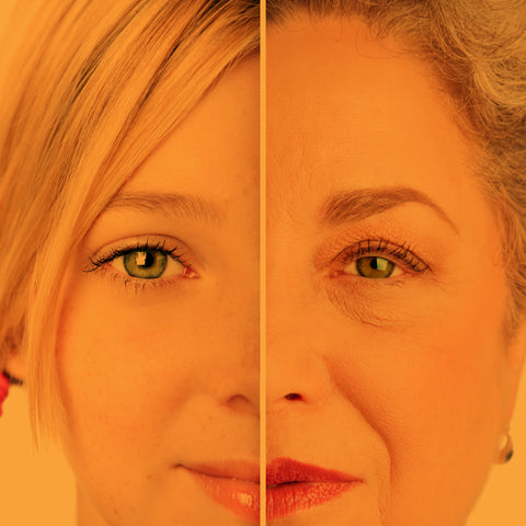
More than a decade ago, a pathbreaking discovery showed for the first time that mature mouse somatic cells could be reprogrammed into pluripotent stem cells (i.e., make them young again).
The discovery was made by using the forced expression of four transcription factors: Oct4, Sox2, Klf4, and c-Myc.
These results came from the research group of Shinya Yamanaka, and hence these factors responsible for reprogramming induced pluripotent stem cells (iPSC) are called the Yamanaka factors [1].
Historical Developments
The generation of iPSC from mature somatic cells signifies a paradigm shift in cellular differentiation. Previously, cellular differentiation was thought to be unidirectional, meaning that pluripotent cells mature into specialized fates, such as muscle, heart cells, and neurons. This led to a strong view in the 19th and early 20th century that mature cells are permanently protected in a differentiated state and unable to revert to a fully immature, pluripotent stem cell [2]. It was thought to be impossible for mature cells to “rewind the clock."
Let’s go through the incredible journey of how imprisoned mature somatic cells are able to break free to become immature pluripotent stem cells.
An Impossible Task
British developmental biologist Conrad Hal Waddington proposed the epigenetic landscape theory for the differentiation of cells from their totipotent to fully differentiated state [3]. Waddington used mountains and valleys as a metaphor to explain his theory of the epigenetic landscape for development. Totipotent cells are represented as the marbles on the mountaintop in this landscape. As they differentiate, these marbles trickle down to stable energy valleys. The assumption of the theory was it would be difficult or nearly impossible to revert differentiated mature cells to an undifferentiated state [2-4].
The failed attempt of reprogramming the somatic cell
Briggs and Kings showed that enucleated eggs could support development up to the tadpole stage when an embryonic nucleus was transferred to them. They were, however, unable to obtain a developing embryo when they transplanted nuclei from more differentiated cells to the enucleated egg. This result led them to conclude that mature differentiated nuclei have lost the ability to promote development, as they have undergone irreversible change during differentiation [2,5].
A ray of hope in reprogramming the somatic cell
In a landmark study, Gurdon showed that a small number of tadpoles could be generated when nuclei from tadpole intestinal epithelium were transferred to enucleated eggs [6]. Gurdon made eggs enucleated by exposing them to ultraviolet radiation, destroying DNA in the chromosomes of eggs. Another reason for the successful result was the change of the model organism; in previous works, scientists used the European frog, Rana, whereas, for Gurdon's work, he employed Xenopus, an African frog [6,7]. With this work, Gurdon concluded that matured somatic cell nuclei had the potential to revert to pluripotency.
First mammal clone
Gurdon's work established somatic cell nuclear transfer (SCNT) in animals, but it took more than three decades to develop the first mammal cloned from SCNT. In 1997, Ian Wilmut described the birth of Dolly, the first mammal born after SCNT from matured mammary epithelial cells [8]. The success of SCNT proved that for the development of entire organisms, even matured somatic cells can work as they contain all the required genetic information [2,9]. SCNT has since been used to clone many mammals, including cats, horses, cows, pigs, and camels [10-13].
The research that led to the foundation of Yamanaka factors and the generation of iPSC
Results from SCNT showed that one could successfully reprogram a matured somatic cell nucleus. However, the question remains whether it is possible to reprogram a matured somatic cell to a highly immature state without using an enucleated egg. Many scientists considered this an impossible task or thought it would require a highly complicated methodology to break open a mature somatic cell to a highly immature state [2].
Learning from SCNT
One of the key observations from SCNT was that the factors that can reprogram matured somatic cell nuclei are present in eggs. Another landmark study showed that matured somatic cell nuclei could be reprogrammed with the fusion of embryonic stem (ES) cells [15]. With the generation of human ES cells, there was widespread interest in reprogramming due to its value in regenerative medicine [16].
Identification of 24 factors for reprogramming
With his experience with mouse ES, Shinya Yamanaka decided to reprogram somatic cells back into the embryonic state [17]. Using the expression sequence tag, he identified the factors specifically expressed in ES cells. He identified numerous genes in undifferentiated mouse ES cells and early embryos whose expressions were specific. The group focused on the genes with the highest enrichment. They categorized genes based on previously published reports as ES cell-specific markers and other ES cell-enriched genes as “ES-cell associated transcripts (ECAT).” They confirmed the expression of ECATs with northern blotting [18,19]. Further experimentation identified a total of 24 factors, including Oct3/4, Sox2, c-Myc, Nanog, other ECATs, and Klf4, as candidate reprogramming factors [1].
They developed an ECAT3 knockout mouse embryonic fibroblast (MEF)-based assay method for assessing the role of reprogramming candidates. When they introduced each of the 24 factors individually, they could not see any reprogramming. They then transduced all 24 factors together in MEFs knockout and, after four weeks, obtained cells that were expandable and showed a morphology similar to that of mouse ES cells.
Yamanaka factors to reprogram mouse and human mature somatic cells
Further analysis showed that clones expressed ES cell markers, including Oct3/4, Nanog, E-Ras, Cripto, Dax1, Zfp296, and Fgf4 [1,20]. Next, they studied the roles of individual factors by withdrawing them from the pool of transduced genes. When they removed Oct3/4 or Klf4, no ES cell-like colonies were observed. Only a few ES-like colonies emerged after the removal of Sox2. On removing c-Myc, they observed ES cell-like colonies, but their morphology didn’t resemble ES cells. ES-cell-like colonies or morphology were not significantly impacted when they removed other factors. They finally showed that it is possible to induce pluripotent stem cells from mouse fibroblast using a combination of four transcription factors: Oct3/4, Klf4, Sox2, and c-Myc [1,2,21]. To check whether iPSC had pluripotency, they injected iPS cells subcutaneously in nude mice and obtained tumors. They performed histological analysis and found that iPS cells differentiated into all three germ layers, including neural tissues, cartilage, and columnar epithelium. They were also able to produce adult chimeras from iPS cells and hence concluded that iPS cells are comparable to ES cells in terms of pluripotency [1].
They then used Yamanaka factors to produce iPS cells from human fibroblast within one year of mouse iPS [22]. At the same time, the Thompson group used different factors to produce human iPS [23]. According to Yamanaka, they were able to produce human iPS quickly due to the available information on human ES culture. He said during his Nobel Lecture, “We could have never generated human iPS cells without the previous reports on human ES cells. [21]"
Applications of Yamanaka Factors
One of the key advantages of the Yamanaka factors is its simplicity and reproducibility. This has led to the revolutionizing of stem cell research. Now many labs across the globe are producing iPS cells for studying multiple physiological features of diseases and injuries. The revolution has been fastened with the availability of ready-made kits containing Yamanaka factors to generate iPS in a simplistic manner [24].
iPS cells for spinal cord injury
Scientists used human iPS cell-derived neurospheres (hiPSC-NS) to treat mouse spinal cord injuries [25]. They observed synapse formation between hiPSC-NS-derived neurons and host mouse neurons. They also observed the expression of neurotrophic factors, angiogenesis, axonal regrowth, and increased amounts of myelin in the injured area. The mouse treated with hiPSC-NS had persistent recovery throughout the study period, significantly better than that of the control mouse. The study found no tumor formation in the mice after transplanting hiPSC-NS, suggesting they are a potential therapeutic source for treatment [9,25].
iPS cells for neurodegenerative disease model
Parkinson's Disease (PD) involves the selective loss of dopaminergic neurons. The progression of the disease and its molecular mechanisms are not yet well-understood. With the advent of iPSC models of PD (iPSC-PD), new revelations are coming up [26]. A study with iPSC-PD showed that neuronal death in PD is induced by oxidative stress caused by mitochondria [27]. Another study showed that aggregation of α-synuclein and Lewy body-like deposition in the dopaminergic neuron is associated with PD [28]. Furthermore, iPSC-PD models generated from PD patients are becoming an important source of preclinical models to understand the pathophysiology of PD [29].
Alzheimer’s Disease (AD) is characterized by brain volume reduction, amyloid-beta protein plaques, and aggregation of tau-proteins leading to neurodegeneration [30]. iPSC-derived neurons from different patients of AD (iPSC-AD) showed different accumulations of amyloid-beta protein [31]. iPSC-AD also showed impaired mitochondrial energy metabolism and reactive oxygen species as the main feature of AD [30-32].
Using iPSC models for AD and PD will pave the future of understanding their pathophysiology, new therapeutics, disease prevention, and usher in the era of novel drug targets to cure the disease [26].
iPSC in other diseases
Cancer: iPSC-derived models are widely used in understanding how cancer starts. Recent studies on chemoresistance in liver cancer have employed the Yamanaka factors [33].
Cardiovascular disease: iPSC-derived cardiomyocytes (iPSC-CM) from patients are becoming a go-to approach to screen new therapies and test drug efficacy. iPSC-CM is the future of personalized medicine [34].
Clinical trials using iPSC
With the advancement of iPSCs in regenerative medicine, they are now being widely used for clinical trials. The first documented clinical trial involving iPSC-derived cells was designed to target a disease that affects the eye's macula, leading to the blurring of central vision [35]. The study observed that patient vision improved after six months of transplantation with iPSC-derived cells. There were no safety-related concerns [35].
With the success of pre-clinical PD studies with iPSC, Kyoto university announced a clinical trial of iPSC-derived dopaminergic progenitors to be transplanted on 7 PD patients [36]. The objective of the Kyoto trial is to evaluate the safety and efficacy of transplanted iPSC into the patient's brain [36]. A recent review documented that there are a total of 81 clinical trials registered with iPSC [37]. Out of these 81, 62 are non-therapeutic and the rest are therapeutic [37].
The Future for Yamanaka Factors
In a very short period of time, the discovery of the Yamanaka factors has transformed regenerative medicine and developmental biology. Applying iPSC in deciphering the complex etiology and pathophysiology of various diseases will help novel drug development.
In the coming decades, significant challenges in the field of iPSC research need to be overcome. These challenges include improving the efficiency of iPSC generation, biobanking of iPSC, development of an allogenic approach for standard treatment, and better markers to predict in vivo therapeutic efficacy and authenticity of grafted cells [38-40]. Nobel Laureate Yamanaka said on the future of iPSC, “I imagine iPSC-based therapies will be readily available for some diseases. I also hope to see several pharmaceuticals that are developed by using iPSC technology put on the market. With luck, maybe iPSC technology will create new approaches to cure cancers and immunological diseases. [38]"
Alongside diagnosing and treating disease, there is also no doubt that iPSCs will be implemented to reverse age and prevent disease as well. Stay tuned! This field is really just getting started!
References
- Takahashi, K.; Yamanaka, S. Induction of Pluripotent Stem Cells from Mouse Embryonic and Adult Fibroblast Cultures by Defined Factors. Cell 2006, 126, 663–676, doi:10.1016/j.cell.2006.07.024.
- Frisén, J., U. Lendahl, and T.P. Mature Cells Can Be Reporgrammed to Become Pluripotent: The 2012 Nobel Prize in Physiology or Medicine–Advanced Information. Nobel Lect. 2012, 1–12.
- Waddington, C.H. The Strategy of the Genes: A Discussion of Some Aspects of Theoretical Biology.; Routledge, 1957; ISBN 1315765470.
- Allen, M. Compelled by the Diagram: Thinking through C. H. Waddington’s Epigenetic Landscape. Contemp. Hist. Presence Vis. Cult. 2015, 4, 119–142, doi:10.5195/contemp.2015.143.
- King, T.J.; Briggs, R. CHANGES IN THE NUCLEI OF DIFFERENTIATING GASTRULA CELLS, AS DEMONSTRATED BY NUCLEAR TRANSPLANTATION. Proc. Natl. Acad. Sci. U. S. A. 1955, 41, 321–325, doi:10.1073/pnas.41.5.321.
- Gurdon, J.B. The Developmental Capacity of Nuclei Taken from Intestinal Epithelium Cells of Feeding Tadpoles. 1962.
- Gurdon, J.B. The Egg and the Nucleus: A Battle for Supremacy (Nobel Lecture). Angew. Chemie - Int. Ed. 2013, 52, 13890–13899, doi:10.1002/anie.201306722.
- Wilmut, I.; Schnieke, A.E.; McWhir, J.; Kind, A.J.; Campbell, K.H.S. Viable Offspring Derived from Fetal and Adult Mammalian Cells. Nature 1997, 385, 810–813.
- Yamanaka, S. Induced Pluripotent Stem Cells: Past, Present, and Future. Cell Stem Cell 2012, 10, 678–684, doi:10.1016/j.stem.2012.05.005.
- Galli, C.; Lagutina, I.; Crotti, G.; Colleoni, S.; Turini, P.; Ponderato, N.; Duchi, R.; Lazzari, G. A Cloned Horse Born to Its Dam Twin. Nature 2003, 424, 635.
- Tian, X.C.; Kubota, C.; Enright, B.; Yang, X. Cloning Animals by Somatic Cell Nuclear Transfer–Biological Factors. Reprod. Biol. Endocrinol. 2003, 1, 1–7.
- Shin, T.; Kraemer, D.; Pryor, J.; Liu, L.; Rugila, J.; Howe, L.; Buck, S.; Murphy, K.; Lyons, L.; Westhusin, M. A Cat Cloned by Nuclear Transplantation. Nature 2002, 415, 859.
- Keefer, C.L. Artificial Cloning of Domestic Animals. Proc. Natl. Acad. Sci. 2015, 112, 8874–8878.
- Pan, G.; Wang, T.; Yao, H.; Pei, D. Somatic Cell Reprogramming for Regenerative Medicine: SCNT vs. IPS Cells. Bioessays 2012, 34, 472–476.
- Tada, M.; Takahama, Y.; Abe, K.; Nakatsuji, N.; Tada, T. Nuclear Reprogramming of Somatic Cells by in Vitro Hybridization with ES Cells. Curr. Biol. 2001, 11, 1553–1558.
- Thomson, J.A.; Itskovitz-Eldor, J.; Shapiro, S.S.; Waknitz, M.A.; Swiergiel, J.J.; Marshall, V.S.; Jones, J.M. Embryonic Stem Cell Lines Derived from Human Blastocysts. Science (80-. ). 1998, 282, 1145–1147.
- Yamanaka, S.; Zhang, X.-Y.; Maeda, M.; Miura, K.; Wang, S.; Farese, R. V; Iwao, H.; Innerarity, T.L. Essential Role of NAT1/P97/DAP5 in Embryonic Differentiation and the Retinoic Acid Pathway. EMBO J. 2000, 19, 5533–5541.
- Tokuzawa, Y.; Kaiho, E.; Maruyama, M.; Takahashi, K.; Mitsui, K.; Maeda, M.; Niwa, H.; Yamanaka, S. Fbx15 Is a Novel Target of Oct3/4 but Is Dispensable for Embryonic Stem Cell Self-Renewal and Mouse Development. Mol. Cell. Biol. 2003, 23, 2699–2708.
- Takahashi, K.; Mitsui, K.; Yamanaka, S. Role of ERas in Promoting Tumour-like Properties in Mouse Embryonic Stem Cells. Nature 2003, 423, 541–545.
- Mitsui, K.; Tokuzawa, Y.; Itoh, H.; Segawa, K.; Murakami, M.; Takahashi, K.; Maruyama, M.; Maeda, M.; Yamanaka, S. The Homeoprotein Nanog Is Required for Maintenance of Pluripotency in Mouse Epiblast and ES Cells. Cell 2003, 113, 631–642.
- Yamanaka, S. The Winding Road to Pluripotency (Nobel Lecture). Angew. Chemie - Int. Ed. 2013, 52, 13900–13909, doi:10.1002/anie.201306721.
- Takahashi, K.; Tanabe, K.; Ohnuki, M.; Narita, M.; Ichisaka, T.; Tomoda, K.; Yamanaka, S. Induction of Pluripotent Stem Cells from Adult Human Fibroblasts by Defined Factors. Cell 2007, 131, 861–872, doi:10.1016/j.cell.2007.11.019.
- Yu, J.; Vodyanik, M.A.; Smuga-Otto, K.; Antosiewicz-Bourget, J.; Frane, J.L.; Tian, S.; Nie, J.; Jonsdottir, G.A.; Ruotti, V.; Stewart, R.; et al. Induced Pluripotent Stem Cell Lines Derived from Human Somatic Cells. Science (80-. ). 2007, 318, 1917–1920, doi:10.1126/science.1151526.
- Guide, U. CTS TM CytoTune TM -IPS Sendai 2.1 Reprogramming Kit. 1–68.
- Nori, S.; Okada, Y.; Yasuda, A.; Tsuji, O.; Takahashi, Y.; Kobayashi, Y.; Fujiyoshi, K.; Koike, M.; Uchiyama, Y.; Ikeda, E.; et al. Grafted Human-Induced Pluripotent Stem-Cell-Derived Neurospheres Promote Motor Functional Recovery after Spinal Cord Injury in Mice. Proc. Natl. Acad. Sci. U. S. A. 2011, 108, 16825–16830, doi:10.1073/pnas.1108077108.
- Valadez-Barba, V.; Cota-Coronado, A.; Hernández-Pérez, O.R.; Lugo-Fabres, P.H.; Padilla-Camberos, E.; Díaz, N.F.; Díaz-Martínez, N.E. IPSC for Modeling Neurodegenerative Disorders. Regen. Ther. 2020, 15, 332–339, doi:10.1016/j.reth.2020.11.006.
- Ryan, S.D.; Dolatabadi, N.; Chan, S.F.; Zhang, X.; Akhtar, M.W.; Parker, J.; Soldner, F.; Sunico, C.R.; Nagar, S.; Talantova, M. Isogenic Human IPSC Parkinson’s Model Shows Nitrosative Stress-Induced Dysfunction in MEF2-PGC1α Transcription. Cell 2013, 155, 1351–1364.
- Cooper, O.; Seo, H.; Andrabi, S.; Guardia-Laguarta, C.; Graziotto, J.; Sundberg, M.; McLean, J.R.; Carrillo-Reid, L.; Xie, Z.; Osborn, T. Pharmacological Rescue of Mitochondrial Deficits in IPSC-Derived Neural Cells from Patients with Familial Parkinson’s Disease. Sci. Transl. Med. 2012, 4, 141ra90-141ra90.
- Smith, H.L.; Freeman, O.J.; Butcher, A.J.; Holmqvist, S.; Humoud, I.; Schätzl, T.; Hughes, D.T.; Verity, N.C.; Swinden, D.P.; Hayes, J. Astrocyte Unfolded Protein Response Induces a Specific Reactivity State That Causes Non-Cell-Autonomous Neuronal Degeneration. Neuron 2020, 105, 855–866.
- Demetrius, L.A.; Driver, J. Alzheimer’s as a Metabolic Disease. Biogerontology 2013, 14, 641–649.
- Kondo, T.; Asai, M.; Tsukita, K.; Kutoku, Y.; Ohsawa, Y.; Sunada, Y.; Imamura, K.; Egawa, N.; Yahata, N.; Okita, K. Modeling Alzheimer’s Disease with IPSCs Reveals Stress Phenotypes Associated with Intracellular Aβ and Differential Drug Responsiveness. Cell Stem Cell 2013, 12, 487–496.
- Israel, M.A.; Yuan, S.H.; Bardy, C.; Reyna, S.M.; Mu, Y.; Herrera, C.; Hefferan, M.P.; Van Gorp, S.; Nazor, K.L.; Boscolo, F.S. Probing Sporadic and Familial Alzheimer’s Disease Using Induced Pluripotent Stem Cells. Nature 2012, 482, 216–220.
- Fatma, H.; Siddique, H.R. Pluripotency Inducing Yamanaka Factors: Role in Stemness and Chemoresistance of Liver Cancer. Expert Rev. Anticancer Ther. 2021, 21, 853–864, doi:10.1080/14737140.2021.1915137.
- Thomas, D.; Cunningham, N.J.; Shenoy, S.; Wu, J.C. Human-Induced Pluripotent Stem Cells in Cardiovascular Research: Current Approaches in Cardiac Differentiation, Maturation Strategies, and Scalable Production. Cardiovasc. Res. 2022, 118, 20–36, doi:10.1093/cvr/cvab115.
- Bragança, J.; Lopes, J.A.; Mendes-Silva, L.; Santos, J.M.A. Induced Pluripotent Stem Cells, a Giant Leap for Mankind Therapeutic Applications. World J. Stem Cells 2019, 11, 421–430, doi:10.4252/wjsc.v11.i7.421.
- Takahashi, J. IPS Cell-Based Therapy for Parkinson’s Disease: A Kyoto Trial. Regen. Ther. 2020, 13, 18–22.
- Kim, J.Y.; Nam, Y.; Rim, Y.A.; Ju, J.H. Review of the Current Trends in Clinical Trials Involving Induced Pluripotent Stem Cells. Stem cell Rev. reports 2022, 18, 142–154, doi:10.1007/s12015-021-10262-3.


Comments (0)
There are no comments for this article. Be the first one to leave a message!