
Neuronal communication is kind of a big deal (#understatement) and there are two main mechanisms.
One is fast communication, where point-to-point information transfer is mediated by synaptic transmission. This typically occurs on the order of 1/1000 of a second or less than one millisecond. That is really, really fast. The other slow, more widespread signaling is mediated by a range of messengers, such as neuropeptides, endocannabinoids, and monoamines [2]. Slow transmission occurs over hundreds of milliseconds to minutes. Neurons use both mechanisms to transfer information from one neuron to the next and send signals from the brain to other cells in the body [3].
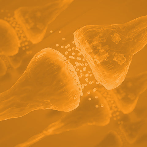
What is synaptogenesis?
The process of forming new synapses between neurons in the brain is called synaptogenesis [4]. Studies have found that synaptogenesis plays a critical role in brain development and plasticity and is believed to underlie many aspects of learning and memory [5-8]. The process of synaptogenesis occurs throughout the lifetime of an organism although it is most prominent during the early development of the nervous system.
During the initial phase of synapse formation, presynaptic and postsynaptic membranes have to be identified and matched, and initial contact must be formed and stabilized [2,4].
What is the best definition of synaptogenesis?
Synaptogenesis is the formation of synapses between neurons within the central nervous system, a fundamental process for establishing and refining neural circuits. It involves complex cellular and molecular mechanisms, including excitatory synaptogenesis and the interaction of synaptic vesicle proteins, crucial for cognitive development and neuronal activity across the lifespan.
The Discovery of the Synapse
Early to late 19th Century
Synapses -- the concept of the action potential with its action current showing up as a negative variation -- were first demonstrated by Emil duBois-Reymond in 1842 [9]. Cajal and Golgi discovered how nervous systems are structured and function [9]. In 1892, Cajal showed how a synapse looked morphologically. In 1897, the term "synapse" was coined by Michael Foster [2]. Golgi and Cajal received the Nobel Prize in Medicine for their discovery in 1906 [10].
20th Century
Although DuBois-Reymond already suggested the chemical nature of synaptic transmission, it was long disputed because of its incredible speed. Over time, however, landmark studies by Katz, von Euler, and Axelord established overwhelming evidence that most synapses use chemical messengers called neurotransmitters [2]. Katz, von Euler, and Axelord were awarded the Nobel Prize in 1970 for discovering neurotransmitter release at the nerve terminal.
21st Century
Even though it was clear that most synapses use neurotransmitters, the molecular mechanism of their release was not understood until the late 1990s. The groundbreaking work of Thomas Südhof focused on the molecular mechanisms of neurotransmitter release. This is the first step of synaptic transmission that accounts for the brain's speed and precision of information transfer. Südhof received the Nobel Prize for elucidating this mechanism in 2013.
Mechanisms of Synaptogenesis
There is a much better understanding of the organization of the core functions of the synapse due to extensive research carried out by scientists. However, the current molecular understanding of how a synapse is formed and maintained is not completely understood. Three approaches have been proposed below.
Transmembrane proteins involvement in synapse formation
Studies have found that a diverse set of membrane proteins and pathways are used by neurons to control and initiate synapse formation. The commonly involved transmembrane proteins in synapse development include:
- Synaptic Cell Adhesion Molecules (SynCAMs) [11]
- Cadherins [12]
- Teneurins [13]
- Epidermal Growth Factor (EGF) repeats containing transmembrane proteins [14]
- Neurexin-Neuroligin pairs [15]
- Leucine-rich-repeat kinase (LRRK) [16]
- Eph–ephrins [17]
Many early synaptogenic proteins have additional axon branching and pathfinding roles. Using these synaptic proteins, studies have found that one presynaptic neuron could even form different synapse types with different postsynaptic target cells.
Neurotrophic factors
The neurotrophin brain-derived neurotrophic factor (BDNF) regulates synaptogenesis by its signaling pathways via the tropomyosin receptor kinase B (TrkB) [18-20]. BDNF and TrkB are highly expressed in areas of the brain involved in learning and memory. BDNF regulates synaptogenesis in three ways [21]:
- Increasing the arborization of axons and dendrites
- Inducing axonal and dendritic bouton formation
- Stabilizing existing synapses
To show that BDNF affects synaptogenesis in the developmental stage, a mouse study with loss-of-function human variants of BDNF-TrkB found that BDNF-stimulated synaptogenesis was affected, particularly in the maturation of functional synapses [19]. With scientific knowledge available on the mechanism of new synapse assembly, it can now be said with certainty that TrkB signaling, and BDNF specifically, modulates synaptogenesis in the “critical period” of development and adulthood [18,19,21].
Gene expression
The formation of new synapses also requires the correct expression of specific genes. Studies have shown that the activity-dependent regulation of gene expression is necessary for synaptogenesis [2,16]. These genes include [2,3,16,22-26]:
- Neurexin
- Neuroligin
- Shank
- Synaptotagmin
- Synaptophysin
- Munc13
- Synapsin
What happens in synaptogenesis?
During synaptogenesis, neurons form new synaptic connections, involving the growth of axons and dendrites towards each other. This process is guided by cellular and molecular mechanisms, including the role of neurotransmitters like glutamate, and structures such as growth cones, leading to changes in synaptic density and enhanced synaptic connectivity in the nervous system.
At what age does synaptogenesis occur?
Synaptogenesis occurs throughout a person's lifespan but is most pronounced during early childhood and adolescence. This phase is crucial for brain development, especially in the cerebral cortex, where a surge in synaptic connectivity supports learning and memory. In the adult brain, synaptogenesis continues, albeit at a slower pace, contributing to neural plasticity.
Diseases associated with the dysfunction of synaptogenesis
Many diseases have been reported to be associated with the failure of synaptogenesis, leading to the loss of connections between neurons. Some of the most common include:
- Autism is a neurological developmental disorder characterized by impaired glutamatergic synapse formation, development, and maintenance. Several studies found that genetic alterations in Shank and Neuroligin have been associated with autism [27-32].
- Schizophrenia (SCZ) is a neurological disorder classified by delusions, hallucinations, and disorganized behavior that results in social or occupational dysfunction [33,34]. One of the major causes of SCZ is impaired glutamatergic synapse formation and dysfunctional synaptic transmission. This leads to abnormal connectivity between the prefrontal cortex and the limbic system, striatum, and thalamus [34,35].
- Alzheimer's Disease (AD) is a progressive neurodegenerative disorder that leads to the loss of cognitive function and memory [32]. Studies have shown that the amyloid plaques and neurofibrillary tangles that form in AD can disrupt the normal process of synaptogenesis [36,37]. This can reduce the number and strength of synapses and affect the normal synaptic function.
- Parkinson's Disease (PD) is a neurodegenerative disorder characterized by the loss of dopamine-producing nerve cells in a specific area of the brain [32]. Cortical synaptogenesis may be impaired in the areas of the brain responsible for movement control, leading to the disease's motor symptoms. LRRK has been associated with up to 40% of sporadic PD cases in defined cohorts [32].
5 Ways to Enhance Synaptogenesis
Let's dive into the cellular and molecular mechanisms, exploring how activities ranging from motor learning to dietary adjustments can foster exuberant synaptogenesis.
-
Motor Learning
Acquiring new knowledge is beneficial for the brain, but not all types of learning can lead to the formation of new synapses. To foster the formation of newly formed synapses, it is important to challenge the brain to coordinate and perform complex movements in conjunction with a specific task [38]. This could be acrobatic training, for example. Engaging in motor learning, such as learning a new technique in table tennis or juggling, can also lead to the formation of new synapses that help to improve that skill [39].
A study found that adult rats trained in complex acrobatic movements developed new synapses, while rats assigned to simple physical exercise or no activity did not [40]. The study also revealed no significant difference in the synaptic density in the rat brain between the group of rats that exercised and the group that did not, indicating that exercise alone may not be sufficient to promote the formation of new synapses [40].
-
Antioxidants in the Diet
Synaptogenesis can be increased by a higher intake of antioxidants in the diet [41]. A research study examined the role of antioxidants in the effectiveness of supplementing with a combination of uridine and omega-3 fatty acids (docosahexaenoic acid and eicosatetraenoic acid) in rats [42]. The results showed that the levels of phospholipids, the pre- and post-synaptic proteins, were significantly improved by taking a combination supplement of omega-3 fatty acids, uridine, and antioxidants.
The study also observed that a supplement containing omega-3 fatty acids, uridine, vitamin C, vitamin E, and selenium was found to have a strong positive effect on synaptogenesis. However, the supplementation with omega-3 fatty acids and uridine alone had less of an effect, indicating that vitamin C, vitamin E, and selenium play a crucial role in promoting synaptogenesis [41,42].
-
Methylene Blue (our favorite, obviously)
Methylene blue (MB) has been shown to have neuroprotective effects, including the reduction of aggregated proteins, enhancement of antioxidant response, and improvement of mitochondrial function and survival in various models of neurodegenerative diseases [43].
MB has been found to promote the formation of new synapses in the hippocampus, a brain region involved in learning and memory, in animal models. It also increases the production of BDNF, a neurotrophic factor involved in synaptogenesis [43]. A study in rats observed that MB might play a role in creating new neurons by improving neuroinflammation, promoting the growth of neurites, and fostering synaptogenesis [44].
Another study focusing on the role of MB as a neuroprotector in cisplatin-induced neurotoxicity found that MB was associated with the positive regulation of synaptic plasticity [45]. A clinical trial that used a randomized, double-blind, and placebo-controlled design found that administering MB led to an increased response in the bilateral insular cortex during a psychomotor vigilance task and also resulted in a 7% improvement in correct responses when recalling memories [46]. These studies show the importance of MB in improving the formation of new synapses and synaptic plasticity. You can get our MB troche here and learn more about it in several of our blogs including this one.
-
Resveratrol
Resveratrol is a beneficial antioxidant and anti-inflammatory compound found in grapes, red wine, raspberries, and dark chocolate. It is known to prevent the development of neurodegenerative diseases. Researchers now understand that resveratrol can help maintain the integrity of the blood-brain barrier, support the mitochondria, and increase blood flow to the brain [47-49]. Additionally, it has been found to promote and enhance the formation of new synapses [47].
-
Intensive Exercise
In a small-scale human study, after five weeks of aerobic training, improvements were observed in physical fitness, cognitive abilities, and increased BDNF in the blood in response to acute exercise [50]. A randomized controlled trial was conducted on 120 older adults, and it was found that regular aerobic exercise increases the size of the anterior hippocampus, resulting in improved spatial memory [51]. The study revealed that exercise training increased the volume of the hippocampus by 2%, which effectively reversed the age-related loss of volume by one to two years.
Additionally, it was found that the increased hippocampus volume was linked to higher levels of BDNF in the blood, a compound that promotes neurogenesis in the dentate gyrus. In contrast, the control group showed a decline in the volume of the hippocampus. These findings suggest that regular aerobic exercise effectively reverses the loss of hippocampus volume in late adulthood, which is associated with improved memory [51]. These studies suggest that intense exercise increases the BDNF level and might play a role in synaptogenesis.
Outlook
Despite many years of research, synaptogenesis remains an enigma [2-4,16]. Recent advancements have revealed certain molecules and processes that play a crucial role in the formation of synapses. However, a complete understanding of how pre- and post-synaptic neurons connect and form a synapse, as well as the signaling pathways that govern this process, is still lacking [2-4,16]. Many questions still need to be answered to gain a holistic understanding of synapse formation.
It seems quite likely, however, that enhancing synaptogenesis in adults may decrease the incidence of neurodegenerative disorders. So don't wait until it's too late. Get learning, get moving, keep blueing yourself, and increase those antioxidants in your diet!
References
- Hines, M.L.; Carnevale, N.T. NEURON: A Tool for Neuroscientists. Neurosci. 2001, 7, 123–135.
- Südhof, T.C. The Molecular Machinery of Neurotransmitter Release (Nobel Lecture). Angew. Chemie - Int. Ed. 2014, 53, 12696–12717, doi:10.1002/anie.201406359.
- Südhof, T.C. Towards an Understanding of Synapse Formation. Neuron 2018, 100, 276–293, doi:10.1016/j.neuron.2018.09.040.
- Petzoldt, A.G.; Sigrist, S.J. Synaptogenesis. Curr. Biol. 2014, 24, R1076–R1080, doi:10.1016/j.cub.2014.10.024.
- Kelsch, W.; Sim, S.; Lois, C. Watching Synaptogenesis in the Adult Brain. Annu. Rev. Neurosci. 2010, 33, 131–149.
- Chugani, H.T. Biological Basis of Emotions: Brain Systems and Brain Development. New Perspect. early Emot. Dev. Miami Johnson Johnson Pediatr. Inst. 1998, 5–16.
- Kolb, B.; Mychasiuk, R.; Muhammad, A.; Gibb, R. Brain Plasticity in the Developing Brain. Prog. Brain Res. 2013, 207, 35–64.
- Kolb, B.; Gibb, R. Brain Plasticity and Behaviour in the Developing Brain. J. Can. Acad. Child Adolesc. Psychiatry 2011, 20, 265.
- Bennett, M.R. The Early History of the Synapse: From Plato to Sherrington. Brain Res. Bull. 1999, 50, 95–118, doi:10.1016/S0361-9230(99)00094-5.
- Nobel Prize Nobel Prize in Medicine 1906 Available online: https://www.nobelprize.org/prizes/medicine/1906/summary/ (accessed on 12 January 2023).
- Biederer, T.; Sara, Y.; Mozhayeva, M.; Atasoy, D.; Liu, X.; Kavalali, E.T.; Südhof, T.C. SynCAM, a Synaptic Adhesion Molecule That Drives Synapse Assembly. Science (80-. ). 2002, 297, 1525–1531, doi:10.1126/science.1072356.
- Yamagata, M.; Duan, X.; Sanes, J.R. Cadherins Interact With Synaptic Organizers to Promote Synaptic Differentiation . Front. Mol. Neurosci. 2018, 11.
- Zhang, X.; Lin, P.-Y.; Liakath-Ali, K.; Südhof, T.C. Teneurins Assemble into Presynaptic Nanoclusters That Promote Synapse Formation via Post-synaptic Non-Teneurin Ligands. Nat. Commun. 2022, 13, 2297, doi:10.1038/s41467-022-29751-1.
- Haltom, A.R.; Jafar-Nejad, H. The Multiple Roles of Epidermal Growth Factor Repeat O-Glycans in Animal Development. Glycobiology 2015, 25, 1027–1042, doi:10.1093/glycob/cwv052.
- Craig, A.M.; Kang, Y. Neurexin-Neuroligin Signaling in Synapse Development. Curr. Opin. Neurobiol. 2007, 17, 43–52, doi:10.1016/j.conb.2007.01.011.
- Südhof, T.C. The Cell Biology of Synapse Formation. J. Cell Biol. 2021, 220, 1–18, doi:10.1083/jcb.202103052.
- Kania, A.; Klein, R. Mechanisms of Ephrin–Eph Signalling in Development, Physiology and Disease. Nat. Rev. Mol. Cell Biol. 2016, 17, 240–256, doi:10.1038/nrm.2015.16.
- Park, H.; Poo, M. Neurotrophin Regulation of Neural Circuit Development and Function. Nat. Rev. Neurosci. 2013, 14, 7–23.
- Sonoyama, T.; Stadler, L.K.J.; Zhu, M.; Keogh, J.M.; Henning, E.; Hisama, F.; Kirwan, P.; Jura, M.; Blaszczyk, B.K.; DeWitt, D.C. Human BDNF/TrkB Variants Impair Hippocampal Synaptogenesis and Associate with Neurobehavioural Abnormalities. Sci. Rep. 2020, 10, 1–14.
- Luikart, B.W.; Nef, S.; Virmani, T.; Lush, M.E.; Liu, Y.; Kavalali, E.T.; Parada, L.F. TrkB Has a Cell-Autonomous Role in the Establishment of Hippocampal Schaffer Collateral Synapses. J. Neurosci. 2005, 25, 3774–3786.
- Wang, C.S.; Kavalali, E.T.; Monteggia, L.M. BDNF Signaling in Context: From Synaptic Regulation to Psychiatric Disorders. Cell 2022, 185, 62–76, doi:10.1016/j.cell.2021.12.003.
- Dean, C.; Dresbach, T. Neuroligins and Neurexins: Linking Cell Adhesion, Synapse Formation and Cognitive Function. Trends Neurosci. 2006, 29, 21–29.
- Guilmatre, A.; Huguet, G.; Delorme, R.; Bourgeron, T. The Emerging Role of SHANK Genes in Neuropsychiatric Disorders. Dev. Neurobiol. 2014, 74, 113–122.
- Lipstein, N.; Chang, S.; Lin, K.-H.; López-Murcia, F.J.; Neher, E.; Taschenberger, H.; Brose, N. Munc13-1 Is a Ca2+-Phospholipid-Dependent Vesicle Priming Hub That Shapes Synaptic Short-Term Plasticity and Enables Sustained Neurotransmission. Neuron 2021, 109, 3980–4000.
- Geppert, M.; Goda, Y.; Hammer, R.E.; Li, C.; Rosahl, T.W.; Stevens, C.F.; Südhof, T.C. Synaptotagmin I: A Major Ca2+ Sensor for Transmitter Release at a Central Synapse. Cell 1994, 79, 717–727.
- Feany, M.B.; Yee, A.G.; Delvy, M.L.; Buckley, K.M. The Synaptic Vesicle Proteins SV2, Synaptotagmin and Synaptophysin Are Sorted to Separate Cellular Compartments in CHO Fibroblasts. J. Cell Biol. 1993, 123, 575–584.
- Laumonnier, F.; Bonnet-Brilhault, F.; Gomot, M.; Blanc, R.; David, A.; Moizard, M.-P.; Raynaud, M.; Ronce, N.; Lemonnier, E.; Calvas, P.; et al. X-Linked Mental Retardation and Autism Are Associated with a Mutation in the NLGN4 Gene, a Member of the Neuroligin Family. Am. J. Hum. Genet. 2004, 74, 552–557, doi:10.1086/382137.
- Chanda, S.; Marro, S.; Wernig, M.; Südhof, T.C. Neurons Generated by Direct Conversion of Fibroblasts Reproduce Synaptic Phenotype Caused by Autism-Associated Neuroligin-3 Mutation. Proc. Natl. Acad. Sci. U. S. A. 2013, 110, 16622–16627, doi:10.1073/pnas.1316240110.
- Fang, W.-Q.; Chen, W.-W.; Jiang, L.; Liu, K.; Yung, W.-H.; Fu, A.K.Y.; Ip, N.Y. Overproduction of Upper-Layer Neurons in the Neocortex Leads to Autism-like Features in Mice. Cell Rep. 2014, 9, 1635–1643.
- Jamain, S.; Quach, H.; Betancur, C.; Råstam, M.; Colineaux, C.; Gillberg, I.C.; Soderstrom, H.; Giros, B.; Leboyer, M.; Gillberg, C.; et al. Mutations of the X-Linked Genes Encoding Neuroligins NLGN3 and NLGN4 Are Associated with Autism. Nat. Genet. 2003, 34, 27–29, doi:10.1038/ng1136.
- Hung, A.Y.; Futai, K.; Sala, C.; Valtschanoff, J.G.; Ryu, J.; Woodworth, M.A.; Kidd, F.L.; Sung, C.C.; Miyakawa, T.; Bear, M.F.; et al. Smaller Dendritic Spines, Weaker Synaptic Transmission, but Enhanced Spatial Learning in Mice Lacking Shank1. J. Neurosci. Off. J. Soc Neurosci. 2008, 28, 1697–1708, doi:10.1523/JNEUROSCI.3032-07.2008.
- Lepeta, K.; Lourenco, M. V; Schweitzer, B.C.; Martino Adami, P. V; Banerjee, P.; Catuara-Solarz, S.; de La Fuente Revenga, M.; Guillem, A.M.; Haidar, M.; Ijomone, O.M.; et al. Synaptopathies: Synaptic Dysfunction in Neurological Disorders - A Review from Students to Students. J. Neurochem. 2016, 138, 785–805, doi:10.1111/jnc.13713.
- Tandon, R.; Gaebel, W.; Barch, D.M.; Bustillo, J.; Gur, R.E.; Heckers, S.; Malaspina, D.; Owen, M.J.; Schultz, S.; Tsuang, M. Definition and Description of Schizophrenia in the DSM-5. Schizophr. Res. 2013, 150, 3–10.
- Habela, C.W.; Song, H.; Ming, G.-L. Modeling Synaptogenesis in Schizophrenia and Autism Using Human IPSC Derived Neurons. Mol. Cell. Neurosci. 2016, 73, 52–62, doi:10.1016/j.mcn.2015.12.002.
- Mirnics, K.; Middleton, F.A.; Lewis, D.A.; Levitt, P. Analysis of Complex Brain Disorders with Gene Expression Microarrays: Schizophrenia as a Disease of the Synapse. Trends Neurosci. 2001, 24, 479–486.
- Parihar, M.S.; Brewer, G.J. Amyloid-β as a Modulator of Synaptic Plasticity. J. Alzheimers. Dis. 2010, 22, 741–763, doi:10.3233/JAD-2010-101020.
- Marsh, J.; Alifragis, P. Synaptic Dysfunction in Alzheimer’s Disease: The Effects of Amyloid Beta on Synaptic Vesicle Dynamics as a Novel Target for Therapeutic Intervention. Neural Regen. Res. 2018, 13, 616–623, doi:10.4103/1673-5374.230276.
- Kleim, J.A.; Barbay, S.; Cooper, N.R.; Hogg, T.M.; Reidel, C.N.; Remple, M.S.; Nudo, R.J. Motor Learning-Dependent Synaptogenesis Is Localized to Functionally Reorganized Motor Cortex. Neurobiol. Learn. Mem. 2002, 77, 63–77.
- Yamasaki, T. Benefits of Table Tennis for Brain Health Maintenance and Prevention of Dementia. Encyclopedia 2022, 2, 1577–1589.
- Tamakoshi, K.; Ishida, A.; Takamatsu, Y.; Hamakawa, M.; Nakashima, H.; Shimada, H.; Ishida, K. Motor Skills Training Promotes Motor Functional Recovery and Induces Synaptogenesis in the Motor Cortex and Striatum after Intracerebral Hemorrhage in Rats. Behav. Brain Res. 2014, 260, 34–43.
- Varesi, A.; Campagnoli, L.I.M.; Carrara, A.; Pola, I.; Floris, E.; Ricevuti, G.; Chirumbolo, S.; Pascale, A. Non-Enzymatic Antioxidants against Alzheimer’s Disease: Prevention, Diagnosis and Therapy. Antioxidants 2023, 12, 180.
- Cansev, M.; Turkyilmaz, M.; Sijben, J.W.C.; Sevinc, C.; Broersen, L.M.; van Wijk, N. Synaptic Membrane Synthesis in Rats Depends on Dietary Sufficiency of Vitamin C, Vitamin E, and Selenium: Relevance for Alzheimer’s Disease. J. Alzheimers. Dis. 2017, 59, 301–311, doi:10.3233/JAD-170081.
- Bhurtel, S.; Katila, N.; Neupane, S.; Srivastav, S.; Park, P.; Choi, D. Methylene Blue Protects Dopaminergic Neurons against MPTP‐induced Neurotoxicity by Upregulating Brain‐derived Neurotrophic Factor. Ann. N. Y. Acad. Sci. 2018, 1431, 58–71.
- Ahmed, M.E.; Tucker, D.; Dong, Y.; Lu, Y.; Zhao, N.; Wang, R.; Zhang, Q. Methylene Blue Promotes Cortical Neurogenesis and Ameliorates Behavioral Deficit after Photothrombotic Stroke in Rats. Neuroscience 2016, 336, 39–48, doi:10.1016/j.neuroscience.2016.08.036.
- Krutskikh, E.P.; Potanina, D. V.; Samoilova, N.A.; Gryaznova, M. V.; Sadovnikova, I.S.; Gureev, A.P.; Popov, V.N. Brain Protection by Methylene Blue and Its Derivative, Azur B, via Activation of the Nrf2/ARE Pathway in Cisplatin-Induced Cognitive Impairment. Pharmaceuticals 2022, 15.
- Rodriguez, P.; Zhou, W.; Barrett, D.W.; Altmeyer, W.; Gutierrez, J.E.; Li, J.; Lancaster, J.L.; Gonzalez-Lima, F.; Duong, T.Q. Multimodal Randomized Functional MR Imaging of the Effects of Methylene Blue in the Human Brain. Radiology 2016, 281, 516.
- Tang, F.; Guo, S.; Liao, H.; Yu, P.; Wang, L.; Song, X.; Chen, J.; Yang, Q. Resveratrol Enhances Neurite Outgrowth and Synaptogenesis via Sonic Hedgehog Signaling Following Oxygen-Glucose Deprivation/Reoxygenation Injury. Cell. Physiol. Biochem. 2017, 43, 852–869.
- Yang, J.; Huang, J.; Shen, C.; Cheng, W.; Yu, P.; Wang, L.; Tang, F.; Guo, S.; Yang, Q.; Zhang, J. Resveratrol Treatment in Different Time-Attenuated Neuronal Apoptosis after Oxygen and Glucose Deprivation/Reoxygenation via Enhancing the Activation of Nrf-2 Signaling Pathway in Vitro. Cell Transplant. 2018, 27, 1789–1797.
- Khan, A.; Jahan, S.; Imtiyaz, Z.; Alshahrani, S.; Antar Makeen, H.; Mohammed Alshehri, B.; Kumar, A.; Arafah, A.; Rehman, M.U. Neuroprotection: Targeting Multiple Pathways by Naturally Occurring Phytochemicals. Biomedicines 2020, 8, 284.
- Griffin, É.W.; Mullally, S.; Foley, C.; Warmington, S.A.; O’Mara, S.M.; Kelly, Á.M. Aerobic Exercise Improves Hippocampal Function and Increases BDNF in the Serum of Young Adult Males. Physiol. Behav. 2011, 104, 934–941.
- Erickson, K.I.; Voss, M.W.; Prakash, R.S.; Basak, C.; Szabo, A.; Chaddock, L.; Kim, J.S.; Heo, S.; Alves, H.; White, S.M.; et al. Exercise Training Increases Size of Hippocampus and Improves Memory. Proc. Natl. Acad. Sci. U. S. A. 2011, 108, 3017–3022, doi:10.1073/pnas.1015950108.


Comments (0)
There are no comments for this article. Be the first one to leave a message!